Regulation of cell cycle progression by cell–cell and cell–matrix forces
Times cited: 200
Uroz, M, Wistorf, S, Serra-Picamal, X, Conte, V, Sales-Pardo, M, Roca-Cusachs, P, Guimera, R, Trepat, X.
Nat. Cell Biol.
20
,
646
-654
(2018).
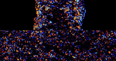
It has long been proposed that the cell cycle is regulated by physical forces at the cell–cell and cell–extracellular matrix (ECM) interfaces. However, the evolution of these forces during the cycle has never been measured in a tissue, and whether this evolution affects cell cycle progression is unknown. Here, we quantified cell–cell tension and cell–ECM traction throughout the complete cycle of a large cell population in a growing epithelium. These measurements unveil temporal mechanical patterns that span the entire cell cycle and regulate its duration, the G1–S transition and mitotic rounding. Cells subjected to higher intercellular tension exhibit a higher probability to transition from G1 to S, as well as shorter G1 and S–G2–M phases. Moreover, we show that tension and mechanical energy are better predictors of the duration of G1 than measured geometric properties. Tension increases during the cell cycle but decreases 3 hours before mitosis. Using optogenetic control of contractility, we show that this tension drop favours mitotic rounding. Our results establish that cell cycle progression is regulated cooperatively by forces between the dividing cell and its neighbours.
Media coverage